


Birth Defects & Fetal Surgery
Pregnancy can be an exciting and smooth journey for women; however, complications may arise and can affect their health, the fetus’s health, or both at the same time.
Dr. Gihad Chalouhi, MD, PhD, Maternal Fetal Medicine – Specialized in Screening and diagnostic fetal ultrasound imaging at Mount Lebanon Hospital (MLH), stresses on the importance of early diagnosis to detect genetic disorders and treat prenatal pathologies. He also explains the two latest techniques in fetal therapy and fetal surgery performed at MLH, after the success of the national Premiere for the treatment of congenital diaphragmatic hernia with plug insertion
https://www.youtube.com/watch?v=YhwPIvCTsz8
How can genetic disorders be diagnosed?
There are several diagnostic invasive procedures performed during pregnancy that can help determine the fetus’ karyotype and identify chromosomal anomalies. These procedures are particularly recommended to parents presenting high risk or who are carriers of a genetic mutation or translocation, this can be explained by a case of previous sibling genetic disorder.
The most common diagnostic tests performed are Chorionic Villus Sampling (CVS) and Amniocentesis (Amniotic Fluid Testing-AFT):
– Chorionic Villus Sampling (CVS)
Commonly performed between weeks 11 and 14 of pregnancy through the extraction of a small sample of placental tissue (chorionic villi). The CVS procedure allows for an early diagnosis of chromosomal anomalies and genetic disorders early diagnosis.
– Amniocentesis or Amniotic Fluid Test (AFT)
Commonly performed starting week 16 of pregnancy through the collection of a small amount of amniotic fluid containing fetal DNA. Amniocentesis allows the examination of fetal DNA to uncover genetic or chromosomal anomalies; in addition, in certain cases to fetal infections.
What is fetal surgery and therapy?
Fetal surgery and therapy are a branch of Maternal and Fetal Medicine (MFM) which allows to treat certain disabling and life-threatening birth defects in the womb, including:
– Congenital Diaphragmatic Hernia
– Fetal Anemia
– Twin-to-Twin Transfusion Syndrome (TTTS)
– TRAP Sequence or Acardiac Twin
– Lower Urinary Tract Obstruction
– Amniotic Band Syndrome
– Fetal Ovarian Cysts
– Arachnoid Cysts
– Spina Bifida
– Several Cardiac Pathologies
What are the latest fetal interventions that took place at Mount Lebanon Hospital?
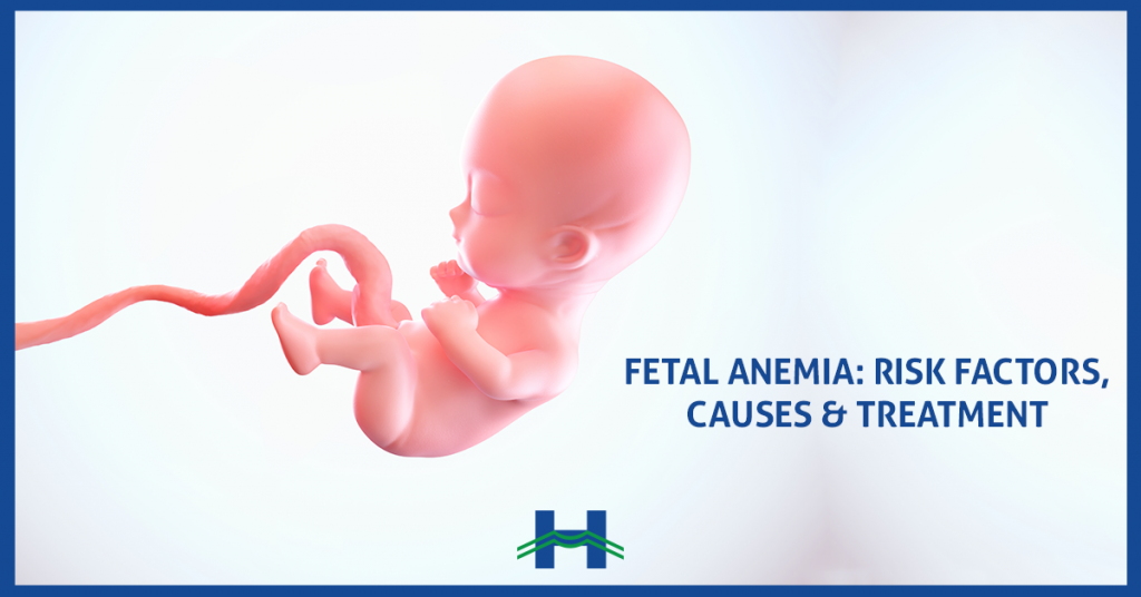
– Fetal Anemia
Fetal anemia occurs mainly due to a Rhesus (Rh) incompatibility between the mother and the fetus’s blood types (the mother is Rh-negative blood and her baby is Rh-positive blood). In this case, the mother’s antibodies cross the placenta to attack and destroy the baby’s red blood cells, the Rh-positive cells. Fetal anemia lowers the ability of the blood to carry oxygen to the baby’s organs and tissues. Depending on the severity, this anemia can become life-threatening for the fetus during pregnancy and after birth.
When diagnosed early, the fetal surgeon performs a blood transfusion of red blood cells (O-negative blood) into the baby’s circulation through the mother’s uterus. The fetus may need more than one transfusion during the pregnancy, depending on when the anemia occurs.
To prevent fetal anemia, pregnant women with Rh-negative blood type should get a preventive injection of antibodies during pregnancy and at delivery (and even in case of miscarriages).
During pregnancy, women with Rh-negative blood type should undergo systematically an indirect Coombs test to screen for antibodies against the Rh factor of the fetus to detect the risk of fetal anemia. In case this test is positive, these women should be followed closely to diagnose fetal anemia when it occurs and treat it in time.
– Twin-to-Twin Transfusion Syndrome
The Twin-to-Twin Transfusion syndrome (TTTS) occurs in monochorionic-diamniotic twin pregnancies (identical twins), where the twins share the same placenta but have separate amniotic sacs. In this condition, the donor twin, transfers significant amounts of blood volume to the other twin, known as the recipient. The donor twin will get less blood volume than its sibling, causing him/her to produce less urine, the main component of amniotic fluid. Meanwhile, the recipient twin will have an overload of blood volume, leading to excess urine production which may lead to heart failure.
When twins are affected by TTTS, the condition is lethal in 90% of cases if not treated. However, the fetal surgeon is able to disconnect the shared blood vessels between the twins in the placenta with the help of a Fetoscopic Laser, to stop the unhealthy transfer between them.
Thanks to the laser coagulation of the vessels with fetoscopy, survival rates for at least one twin becomes greater than 80% and for both twins approximately 50%.
It is thus crucial for pregnant women with monochorionic twins to be closely monitored by a specialist and to undergo an ultrasound every 2 weeks, since TTTS can affect any monochorionic pregnancy in up to 20% of cases (1 in 5 cases).
Can birth defects be prevented?
Some precautions and procedures have proven effective in lowering the risks and preventing birth defects.
Folic acid treatment, starting at least one month before pregnancy, has proven to lower the risk of neural tube defects (congenital anomalies of the nervous system). Daily dose: 400 mcg.
Ultrasound scans are a must during pregnancy. The scan for anomalies will help with the early diagnosis of a wide range of congenital anomalies and birth defects, to be treated in the womb, and allows a better fetal development, which contributes in preparation for a complete and healthy baby at birth.
Dr. Gihad Chalouhi, Fetal Surgeon
Maternal Fetal Medicine Specialist
Screening and diagnostic fetal ultrasound imaging
“A new baby is like the beginning of all things—wonder, hope, a dream of possibilities.” Eda J. LeShan
Leave a reply